Conjoined Twins Abby and Erin Delaney: Inside Their Remarkable Story
In a world where the extraordinary often unfolds in whispered medical journals or behind closed hospital doors, few stories so powerfully capture both the fragility and tenacity of human life as the journey of Abby and Erin Delaney. Born in July 2016 in Philadelphia, Pennsylvania, the two girls entered the world in the rarest of conditions—as craniopagus conjoined twins, fused at the very center of the head. What followed was not just a complex medical odyssey, but a family’s unwavering commitment to hope, innovation, and the promise of a life beyond what was once believed possible.
A Rare Diagnosis, Felt Before Birth
When Heather and Riley Delaney discovered they were expecting twins, it should have been a moment of pure joy. Instead, it quickly became one of profound uncertainty. Around 11 weeks into the pregnancy, doctors revealed that the developing babies were not just twins, but craniopagus conjoined twins—a condition so uncommon it occurs in only a tiny fraction of all twin pregnancies.
For many parents, such a prenatal diagnosis can be shattering. It confronts them with a future filled with unknowns—whether their children will survive birth, whether they can ever live as independent individuals, and what quality of life might be possible if they do. For Heather and Riley, it meant re‑orienting their lives around medical specialists, fetal imaging, and hospital planning long before their daughters arrived.
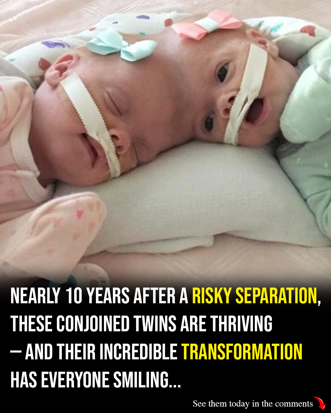
Birth and Immediate Challenges
On July 24, 2016, after a scheduled C‑section delivery, Abby and Erin were born prematurely at around 30 weeks gestation. Weighing barely over two pounds each, they entered the world united at the top of their heads—sharing not only bone and skin but critical vascular connections that supplied blood to each other’s brains.
Premature birth in and of itself presents serious risks, particularly at such early gestational age. But when combined with the complexities of conjoinment, the girls faced a battle for survival from the very first moments of life. They were admitted immediately into the Children’s Hospital of Philadelphia (CHOP) neonatal intensive care unit—a center with world‑class expertise in pediatric care and rare surgical interventions.
For 15 months, the twins would remain in the hospital, watched over by a multidisciplinary team of physicians, nurses, therapists, and support staff. Day after day, their small lives were nurtured, monitored, and charted with painstaking detail.
Preparing for Separation: A Marathon, Not a Sprint
Conjoined twins like Abby and Erin present some of the most intricate surgical challenges in medicine. Doctors must understand exactly how the twins’ brains and blood vessels are connected, whether vital structures are shared, and how separation might impact each child’s neurological development.
For the Delaney twins, the challenge was profound. Not only were they joined at the skull, but their connection extended deep into shared blood vessels and brain membranes. Successfully untangling those connections would require months of preparation, including advanced imaging and surgical planning.
Innovations in medical technology played a key role. Surgeons utilized 3D imaging and computer navigation systems to precisely map the twins’ shared anatomy—particularly the superior sagittal sinus, a major blood vessel that returns blood from the brain to the heart.
This level of pre‑operative planning was essential. Unlike typical surgeries where structures are easier to visualize and separate, these girls were fused in ways that very few surgeons ever encounter in their careers. For the Delaneys’ medical team, it was as much a scientific puzzle as it was a deeply emotional mission.
The 11‑Hour Surgery That Changed Their Lives
The operation itself lasted about 11 hours. It was a high‑wire act of surgical precision: first, delicate shared blood vessels were meticulously isolated and separated; then, the tough protective membrane surrounding their brains—called the dura—was divided; finally, neurosurgeons worked to carefully untangle the shared superior sagittal sinus, the critical vascular bridge between the twins’ circulatory systems.
This was not just surgical separation—it was architectural reconstruction of two tiny, interconnected brains, each with its own right to grow, develop, and exist independently. At several points during the surgery, parts of the medical team worked simultaneously on both girls, carefully orchestrating each move to ensure neither was put at undue risk.
When it was over, the team had done something extraordinary: Abby and Erin were successfully separated. They would now live as two distinct individuals for the first time in their lives.
Continue reading…
